There are five vertebral bodies that make up the lumbar spine better known as the lower back. Nerves exiting off of the spinal cord (at the lower thoracic spine as the spinal cord ends at the upper lumbar spine) travel through the central canal. The central canal is made up of individual rings from each vertebra stacked one on top of the other. The nerves exit one at a time through the sides of these vertebral bodies. Over time, as wear and tear and other degenerative conditions, such as disc degeneration and facet arthritis take their toll on the lower spine, these rings that make up the central canal will begin to narrow. If the narrowing is substantial, it causes compression of the sac of nerves (cauda equina) which will lead to lumbar central (or lumbar spinal) stenosis.
Lumbar spinal stenosis (central stenosis), next to the herniated disc, is one of the most common conditions experienced over time—especially by those who are over 50 years of age with females having the majority of problems. The most common cause of lumbar stenosis is degenerative changes of the spine. The most common problem associated with stenosis is degenerative spondylolysthesis (please see section on that subject in this web site)
Lumbar spinal stenosis can also be linked to other conditions such as tumors, infections, metabolic bone disorders and other known diseases but these are exceedingly rare.
Symptoms
The narrowing of the spinal canal can occasionally cause lower back pain but that is the exception rather than the rule. The most common complaints of lumbar spinal stenosis (central stenosis) are a dull ache or “numbness” in the sacroiliac region and buttocks with prolonged standing or walking that increases with the amount of time doing that activity. The symptoms will start to extend down the back of the legs with prolonged standing or walking. A general weakness or fatigue of the legs and loss of sensation can also occur.
In most cases, standing and bending backwards worsens the symptoms whereas sitting or leaning forward helps to relieve them. As the individual bends forward or crouches, the space within the spinal canal increases and relieves the pressure on the sac of nerves. Generally, symptoms associated with lumbar central stenosis typically worsen over time but many times, they don’t advance and occasionally, they can improve over time even without therapy.
Treatment
Non-Surgical
Anti-inflammatory medications, pain relievers, muscle relaxers and cold/heat therapies can help to relieve lower extremity pain associated with lumbar spinal stenosis. Physical therapy and alternative therapies such as acupuncture are also options. Injections are very helpful to control the symptoms. If all measures have been exhausted without using surgery and pain and symptoms are still occurring, surgery will be the next step to treating the condition.
Surgical
There are surgical procedures that can help relieve the pressure on the nerves and therefore the symptoms of lumbar central stenosis.
- A decompressive laminotomy or laminectomy can be performed to create more space in the spinal canal for the nerves. This is done by removing a portion of the roof of the canal and any spurs that have grown into the canal from the degenerative facets. This is essentially a roto-rooter surgery to make more room for the nerves. Please see section on surgical options for this procedure.
If instability has occurred along with spinal stenosis (example is unstable degenerative spondylolysthesis) spinal fusion stabilization procedure may be required in addition to the decompression surgery. Please see section on surgical options for this procedure.
 |
| This is a top down picture of the effects of aging on the spine. The top picture is a normal spinal canal. The bottom picture shows hypertrophy (enlargement) of the facets in the rear of the canal and bulging of the disc in the front which conspire to narrow the spinal canal. |
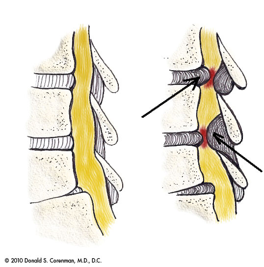 |
| This diagram shows how aging starts to narrow the spinal canal. The arrow in front points to a bulging disc that presses into the canal from front to back. The arrow behind points to ligamentum flavum that has enlarged with aging which pushes into the canal from the rear |
 |
| This illustration demonstrates the change in the diameter in the canal in a patient with spinal stenosis. The picture on the right shows the canal wide open with flexion (bending forward). The picture on the left shows significant compression of the nerve sac in the canal (arrow) with backwards bending. Standing and walking require backwards bending, so the symptoms of central stenosis occur with walking and standing. |
No comments:
Post a Comment