Multiple sclerosis:

The lesions in the deep white matter (yellow arrow) are non specific and can be seen in many diseases.
Typical for MS in this case is:
Typical for MS in this case is:
- Involvement of the temporal lobe (red arrow)
- Juxtacortical lesions (green arrow) - touching the cortex
- Involvement of the corpus callosum (blue arrow)
- Periventricular lesions - touching the ventricles

To search for MS, you must find the following WML in specific sites such as:
-Corpus callosum:

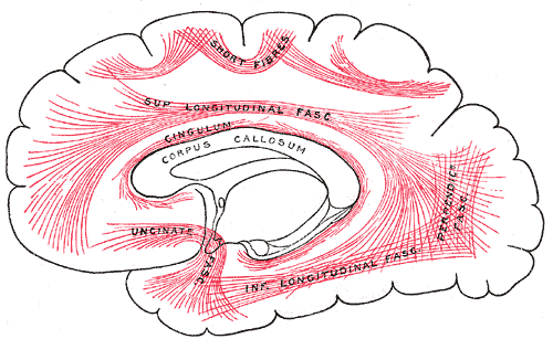
-U-fibers:
The short association fibers (also often referred to as "U-fibers") lie
immediately beneath the gray substance of the cortex of the hemispheres,
and connect together adjacent gyri.
-Infra tentorial:Cerebellum.
- Temporal lobe: early findings.
-Peri ventricular:
-Spinal cord:
-Ga enhancement: yes.
-Dawson's fingers:
-Distribution: Symmetric / diffuse.
Vascular lesions:
-Cortical lesions: Infarction.
-Basal ganglia: Typical.
-Distribution: Assymetric.
So in case of MS; there is many sites of common affection which are:
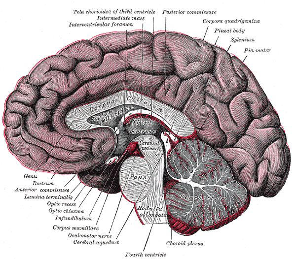
* Corpus callosum.
*Then look above it for U-fibers.
*Then down to temporal lobe.
*then on both sides for peri ventricular areas.
*Then look back and down to cerebellum.
*Then down to spinal cord.
All these lesions are symmetrical.
No comments:
Post a Comment