1-Infarction less than 1 cm in diameter.
2-If lacunar infarction is increased in number with association of ischemic changes in the white matter this leads to formation of a disease known as sub cortical arteriosclerotic encephalopathy.
3-Sub cortical arteriosclerotic encephalopathy disease characterized by diffuse hypo density with multiple lacunar infarction in the white matter and occurs more common in patient with hypertension and diabetes.
4-If you see an infarction with multiple hyper dense spots, looks for the type of infarction if it is acute or chronic, if it is acute, these spots will be hemorrhage, if it is chronic, so these spots will be due to calcification.
 |
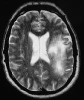
| T2-weighted MRI in a patient infected with HIV
demonstrates a hyperintense lesion in the left frontoparietal region in
the subcortical and periventricular white matter. Biopsy confirmed
progressive multifocal leukodystrophy. |
 |
| Nonenhanced CT of the head shows a hypoattenuating
lesion in the subcortical white matter. Note the characteristic
scalloped lateral margin. |
 |
| Lacunar infarction left basal ganglia |
 |
| Lacunar infarction in T1&T2 |


No comments:
Post a Comment