1-Is due to over production of CSF.
2-Most common cause is choroid plexus papilloma present in the lateral ventricles producing a lot of CSF four or five times the normal range so it can not be drained adequately due to limited capacity of the arachnoid villi.
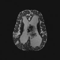 |
| Axial T2-weighted magnetic resonance image (repetition time, 2883 ms; echo time, 100 ms) shows a lobulated mass with frondlike papillary projections in the left lateral ventricle. The mass is isointense relative to the cortex and has internal hypointense foci that likely represent prominent vessels. Note the associated hydrocephalus and transependymal cerebrospinal fluid flow. |
 |
| Interventricular extension through the foramen of Munro, cerebral aqueduct, or foramen of Luschka or Magendie can occur with a choroid plexus papilloma; this is an ancillary diagnostic sign that is not described with other interventricular tumors. |
No comments:
Post a Comment